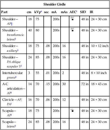
Have you ever wondered how medical professionals determine the precise amount of radiation needed for an X-ray? It’s a crucial aspect of ensuring accurate diagnosis while minimizing potential risks to patients. The answer lies in a critical document known as the X-ray exposure factors chart. These charts act as the “cookbook” for safe and effective X-ray procedures, providing a detailed guide for technicians to ensure optimal image quality with minimal radiation exposure.

Image: gailmorris607o.blogspot.com
This comprehensive guide will delve into the world of X-ray exposure factors charts, explaining their purpose, structure, and significance in the field of medical imaging. We’ll explore how these charts help ensure patient safety, optimize image quality, and ultimately contribute to accurate diagnoses. Whether you’re a healthcare professional seeking to deepen your understanding of these charts or a curious individual interested in learning about the technical aspects of medical imaging, this article will provide valuable insights.
Understanding the Importance of X-Ray Exposure Factors
Every X-ray procedure involves sending a beam of high-energy radiation through the body to create an image. Determining the appropriate amount of radiation to use is essential for producing a clear and diagnostic image while minimizing unnecessary exposure to the patient. This is where X-ray exposure factors come into play.
The primary factors affecting X-ray exposure include:
- kVp (Kilovoltage Peak): This controls the energy of the X-ray beam, influencing the penetration power and image contrast. Higher kVp settings are used for denser body parts, while lower kVp settings are used for less dense tissues.
- mA (Milliamperage): This determines the intensity of the X-ray beam, which directly impacts the amount of radiation hitting the patient. Higher mA settings result in a higher dose but also produce a brighter image.
- Exposure Time: The duration of the X-ray exposure influences the overall radiation dose received by the patient. Shorter exposure times minimize the dose but might compromise image clarity if insufficient radiation reaches the detector.
The Role of X-Ray Exposure Factors Charts
X-ray exposure factors charts act as valuable reference tools for radiographers and technicians, providing a standardized guide for selecting appropriate settings for various imaging procedures. By following these charts, professionals can ensure consistent image quality while minimizing the potential risks associated with radiation exposure. Here are some of the key functions of these charts:
- Standardization: Charts ensure that similar patient types and anatomical regions are imaged consistently, minimizing variations in exposure levels and image quality.
- Optimization: By outlining the optimal exposure factors for specific procedures and anatomical areas, the charts help technicians choose the most effective settings for obtaining clear and diagnostic images.
- Quality Control: Charts provide a reliable benchmark for assessing image quality and ensuring that procedures are performed within acceptable radiation safety standards.
Deconstructing the X-Ray Exposure Factors Chart PDF
X-ray exposure factors charts are typically presented in a PDF format, making them easily accessible and adaptable for various clinical settings. The structure of these charts may vary slightly, but they generally include the following information:
- Anatomical Regions: The charts are organized by anatomical areas, such as chest, abdomen, spine, extremities, etc., allowing for quick and efficient reference.
- Patient Size and Weight: The charts often include specific exposure factors based on the patient’s size and weight, minimizing the need for manual adjustments.
- kVp and mA Settings: The charts outline the recommended kVp and mA settings for each anatomical region, considering factors like bone density, tissue thickness, and desired image contrast.
- Exposure Time: Charts may provide recommended exposure time ranges, depending on the chosen kVp and mA settings and the specific procedure being performed.
- Additional Information: Some charts may include other important information, such as technical details related to the X-ray equipment, specific procedures, and safety guidelines.

Image: www.skylinedentalbend.com
Staying Updated with X-Ray Exposure Factors Charts
The field of medical imaging is constantly evolving, with advancements in technology and best practices influencing radiation safety guidelines. It’s crucial for radiographers to stay updated on the latest recommendations for X-ray exposure factors. Organizations like the **American College of Radiology (ACR)** and the **American Association of Physicists in Medicine (AAPM)** provide valuable resources and guidelines for optimizing imaging procedures.
Here are some ways to stay current with the latest X-ray exposure factors:
- Continuing Education: Participating in continuing education courses and workshops specific to radiation safety and imaging techniques.
- Professional Associations: Joining professional organizations like the ACR and AAPM to access their resources, publications, and webinars on the latest safety standards.
- Manufacturer Guidelines: Regularly reviewing the guidelines and technical specifications provided by the manufacturers of X-ray equipment.
- Online Resources: Exploring reputable online resources, such as the **National Council on Radiation Protection and Measurements (NCRP)** website, for the latest information on radiation safety and imaging practices.
Benefits of Using X-Ray Exposure Factors Charts
The use of X-ray exposure factors charts brings numerous advantages to both patients and healthcare providers.
- Patient Safety: By ensuring appropriate exposure levels, charts contribute to minimizing the radiation dose received by patients, reducing the long-term risks associated with excessive exposure.
- Optimized Image Quality: The standardized settings in these charts optimize image quality by ensuring sufficient radiation reaches the detector while controlling scatter and artifacts, leading to clearer and more diagnostic images.
- Improved Efficiency: By providing a clear roadmap for exposure settings, charts streamline the imaging process, reducing the need for repeated exposures and improving efficiency in the radiology department.
- Enhanced Accuracy: Consistent image quality obtained through the use of charts improves the accuracy of diagnosis and enables more confident decision-making by radiologists.
Examples of X-Ray Exposure Factors Charts
Numerous resources offer readily available X-ray exposure factors charts in PDF format. Some examples include:
- American College of Radiology (ACR): The ACR provides guidelines and resources for radiation safety in medical imaging, including exposure factor charts for various procedures.
- American Association of Physicists in Medicine (AAPM): The AAPM publishes comprehensive resources on radiation safety, dosimetry, and imaging techniques, often featuring exposure factor guidelines.
- National Council on Radiation Protection and Measurements (NCRP): The NCRP develops radiation protection standards and recommendations, which may include exposure factor charts for specific anatomical regions.
- Medical Equipment Manufacturers: X-ray equipment manufacturers provide technical manuals and guidelines for their machines, often including exposure factor charts tailored to their specific devices.
X Ray Exposure Factors Chart Pdf
Conclusion: Embracing the Power of X-Ray Exposure Factors Charts
X-ray exposure factors charts are essential tools for radiographers and technicians, playing a crucial role in delivering safe and effective imaging procedures. These charts streamline the process, optimize image quality, and ensure that patients receive the lowest possible radiation dose for accurate diagnosis. By staying informed about the latest guidelines and adhering to the recommendations within these charts, healthcare professionals can contribute to patient safety, improve diagnostic accuracy, and advance responsible practices in medical imaging. Embrace the power of X-ray exposure factors charts, and you’ll be taking a significant step towards delivering exceptional patient care and ensuring optimal imaging outcomes.


