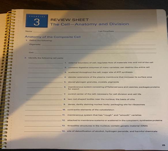
Remember that first time you peered through a microscope, mesmerized by the intricate world within a single cell? It’s a moment that sparks a lifelong fascination with the building blocks of life. While textbooks offer valuable information, hands-on experience through lab exercises is crucial for truly understanding cell anatomy. But what happens when you’re stuck on a lab exercise, struggling to decipher the answers? That’s where this comprehensive guide comes in, providing you with clear, detailed explanations for common Lab Exercise 4 questions on cell anatomy.

Image: studylib.net
This guide isn’t just about providing answers; it’s about demystifying the complex world of cell structure and function. Whether you’re a student encountering cell anatomy for the first time or someone revisiting the subject, this resource will be your trusted companion. We will explore various cell components, their functions, and how they work together to create the intricate machinery of life. So, grab your microscope, get ready to dive deep into the world of cells, and let’s unlock the secrets of Lab Exercise 4!
Exploring the Cellular Landscape
The cell, the fundamental unit of life, is an incredibly complex and organized entity. Its intricate architecture houses a diverse array of organelles, each with a specialized role in maintaining cellular life. Understanding these organelles and their functions is crucial for comprehending the basic processes of life.
The cell membrane, often described as the “gatekeeper,” controls what enters and exits the cell. This thin, flexible barrier is composed primarily of phospholipids and proteins. The nucleus, often referred to as the “command center,” contains the cell’s genetic material, DNA. The DNA provides the instructions for building and maintaining the cell.
Delving Deeper into Lab Exercise 4: Cell Anatomy
Lab Exercise 4 often focuses on identifying and understanding the key organelles within a cell. This exercise typically involves examining prepared slides under a microscope, observing the structures and relating them to their specific functions.
For example, you might be tasked with identifying the mitochondria, often referred to as the “powerhouse of the cell.” These bean-shaped organelles are responsible for cellular respiration, generating energy for the cell’s activities. You’ll also likely encounter the endoplasmic reticulum (ER), a network of interconnected membranes that plays a crucial role in protein synthesis and lipid metabolism. The Golgi apparatus, often envisioned as a “packaging and sorting center,” modifies and packages proteins for secretion.
Dissecting the Secrets of Lab Exercise 4
Here’s a breakdown of common questions that typically appear in Lab Exercise 4, along with detailed explanations and tips for answering them:
- Question: Identify the components of a typical animal cell.
- Answer: A typical animal cell will contain the following organelles: cell membrane, nucleus, cytoplasm, nucleolus, endoplasmic reticulum (ER), Golgi apparatus, ribosomes, mitochondria, lysosomes, and centrioles.
- Tip: Familiarize yourself with the structure and function of each organelle using visual aids like diagrams and videos. Understanding the key role of each component is essential for accurate identification.
A common challenge in cell anatomy lab exercises is distinguishing between plant and animal cells. While both are eukaryotic cells, meaning they have a nucleus and other membrane-bound organelles, they exhibit distinct structural differences.
- Question: Compare and contrast the structure of plant and animal cells.
- Answer: Plant cells are unique, possessing a cell wall composed of cellulose, providing structural support and protection. They also possess chloroplasts, containing chlorophyll for photosynthesis. Animal cells, on the other hand, lack a cell wall and chloroplasts.
- Tip: Practice observing both plant and animal cells under a microscope, paying attention to the key features that differentiate the two. Remember, the presence of a cell wall and chloroplasts signifies a plant cell.
Another common theme in Lab Exercise 4 is the concept of cell division, a fundamental process for growth and repair. Mitosis, a type of cell division, ensures that each daughter cell receives a complete set of chromosomes from the parent cell.
- Question: Describe the stages of mitosis and explain the significance of each stage.
- Answer: Mitosis consists of four distinct phases: prophase, metaphase, anaphase, and telophase. During prophase, the chromosomes condense, making them visible. Metaphase sees the chromosomes align at the center of the cell. Anaphase involves the separation of sister chromatids, moving toward opposite poles of the cell. Lastly, telophase marks the completion of cell division, resulting in two identical daughter cells.
- Tip: Use a visual representation of mitosis, like a diagram or animation, to grasp the sequential steps and understand the significance of each stage. Remember, mitosis is essential for growth, repair, and development.

Image: www.transtutors.com
Unlocking the Cellular Enigma
Beyond the basic cell structures, Lab Exercise 4 might delve into more specialized areas of cell biology. For instance, you might encounter questions about the mechanisms of protein synthesis or the role of cell signaling in communication. These topics require a deeper understanding of cellular processes and the intricate interplay between different organelles.
The key to tackling these more complex questions lies in building a strong foundation of understanding the fundamental principles of cell biology. Visualize the cell as a miniature factory, where each organelle plays a vital role in assembling, modifying, and transporting proteins and other molecules. This perspective helps you connect the dots and appreciate the intricate coordination and communication that occur within a single cell.
FAQ: Answering Your Lab Exercise 4 Queries
Here are answers to some common questions regarding Lab Exercise 4: Cell Anatomy:
- Q: What are the best resources for studying cell anatomy?
A: Textbooks, online tutorials, and interactive simulations are excellent resources. Visualization tools like diagrams, cell animations, and microscopy images can enhance your understanding. - Q: How can I improve my microscope skills for Lab Exercise 4?
A: Practice focusing and manipulating the microscope to obtain clear images. Experiment with different magnifications and lighting settings. Familiarize yourself with the objectives and eyepieces. - Q: What are the common mistakes students make in Lab Exercise 4?
A: Neglecting to label structures correctly, misinterpreting microscope images, and failing to connect the structure and function of identified organelles are common mistakes.
Lab Exercise 4 Cell Anatomy Answers
Empowering Your Cellular Journey
As you embark on your journey through Lab Exercise 4: Cell Anatomy, remember that understanding the intricacies of cell structure and function is not just about memorizing facts; it’s about developing a deeper appreciation for the incredible complexity and beauty of life at its most fundamental level. By carefully observing, analyzing, and interpreting the information from your lab exercises, you’ll unlock a deeper understanding of the living world around us.
Are you excited to delve into the world of cell anatomy and explore the fascinating answers to Lab Exercise 4? Share your thoughts and experiences in the comments below!


